What is a cataract?
A cataract is any cloudiness or opacity of the natural lens of the eye, which is normally crystal clear. There are many types of cataracts. Some cataracts are small and do not interfere with vision. Other cataracts are large and cause severe vision loss.
How common are infantile and childhood cataracts?
Approximately 3 out of 10,000 children have a cataract. The incidence is variable throughout the world.
How does a cataract cause vision loss?
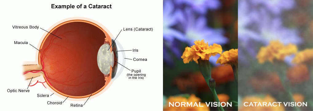
Light enters the eye and is projected to the retina (inner surface of the back of the eye) which senses the light and transmits the signal to the brain. A cataract may stop light from reaching the retina and prevent the eye from seeing. (see figure 2) In order for a child to develop good vision, the child has to have clear light hit the retina and the brain receives a clear image. If there is a cataract blurring the light, it limits the child’s visual development and results in amblyopia. Prompt and sometimes immediate treatment is necessary to prevent permanent vision loss. In contract to adults, cataracts develop after normal visual development so the vision loss can be reversible.
Why are some babies born with a cataract?
Pediatric cataracts often occur because of abnormal lens development during pregnancy. Cataracts can result from genetic problems, infections, or they can occur spontaneously. Lens malformations that occur in conjunction with medical problems are often the result of a genetic or metabolic problem. These cataracts may be present at birth or may develop during childhood. Most pediatric cataracts are isolated findings and are not associated with other abnormalities.
Do all cataracts in babies and children need to be removed?
No. Some cataracts are small and/or off center in the lens. These cataracts do not need to be removed because vision develops normally, even if the cataract is left in place.
How is a cataract removed in infants and children?
A tiny incision is made into the eye and an opening is made in the front of the lens capsule. The very soft and cloudy inner part of the child’s lens is suctioned out of the capsule. Younger children may require an additional opening in the posterior lens capsule with some vitreous gel removal (called a vitrectomy). An intraocular lens (IOL) is then sometimes placed within the empty lens capsule either during the same surgery or in a subsequent second surgery. Generally, an IOL is not placed in a patient less than 1 year of age. Dissolvable stitches are used to close the wounds.